DNA fingerprinting is a method used to identify living things based on samples of their DNA. Instead of looking at the whole sequence of a person’s DNA, these techniques look at the presence or absence of common markers that can be quickly and easily identified.
The process is sometimes called “DNA testing” or “DNA profiling”, but signals the same process. Early DNA fingerprinting was developed in the years before the whole human genome had been sequenced. DNA fingerprinting typically relies on short tandem repeats (STRs), which are unique to individuals. These sections of DNA can be compared between two different samples. If they show the same pattern after gel electrophoresis, it indicates that the samples are from the same source.
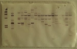
A DNA fingerprint looks something like the columns on the paper below. On this paper, each dark band represents a fragment of VNTRs – and each column is a different tissue sample. A match would be indicated by two columns whose VNTRs patterns matched precisely.

DNA profiling is different from genetic testing, in which a DNA sample is tested to see if it contains genes for inherited diseases or other traits. The FBI and other law enforcement agencies use the CODIS index, which compares 13 sections of DNA and can accurately identify criminals based on a DNA sample.
This process is frequently used in criminal investigations to determine whether blood or tissue samples found at crime scenes could belong to a given suspect. This technology is also used in paternity tests, where comparison of DNA markers can show whether a child could have inherited their markers from the suspected father.
In science, DNA fingerprinting is used in the story of plant and animal populations to determine how closely related species and populations are to other species and populations. Further, it can track their spread over time. This ability to look directly at an organism’s gene markers has revolutionized our understanding of zoology, botany, agriculture, and even human history.
To perform DNA fingerprinting, you must first have a DNA sample! In order to procure this, a sample containing genetic material must be treated with different chemicals. Common sample types used today include blood and cheek swabs.
These samples must be treated with a series of chemicals to break open cell membranes, expose the DNA sample, and remove unwanted components – such as lipids and proteins – until relatively pure DNA emerges.
If the amount of DNA in a sample is small, scientists may wish to perform PCR – Polymerase Chain Reaction – amplification of the sample.
PCR is an ingenious technology which essentially mimics the process of DNA replication carried out by cells. Nucleotides and DNA polymerase enzymes are added, along with “primer” pieces of DNA which will bind to the sample DNA and give the polymerases a starting point.
PCR “cycles” can be repeated until the sample DNA has been copied many times in the lab if necessary.
The best markers for use in quick and easy DNA profiling are those which can be reliably identified using common restriction enzymes, but which vary greatly between individuals.
For this purpose, scientists use repeat sequences – portions of DNA that have the same sequence so they can be identified by the same restriction enzymes, but which repeat a different number of times in different people. Types of repeats used in DNA profiling include Variable Number Tandem Repeats (VNTRs), especially short tandem repeats (STRs), which are also referred to by scientists as “microsatellites” or “minisatellites.”
Once sufficient DNA has been isolated and amplified, if necessary, it must be cut with restriction enzymes to isolate the VNTRs. Restriction enzymes are enzymes that attach to specific DNA sequences and create breaks in the DNA strands.
In genetic engineering, DNA is cut up with restriction enzymes and then “sewn” back together by ligases to create new, recombinant DNA sequences. In DNA profiling, however, only the cutting part is needed. Once the DNA has been cut to isolate the VNTRs, it’s time to run the resulting DNA fragments on a gel to see how long they are!
Gel electrophoresis is a brilliant technology that separates molecules by size. The “gel” in question is a material that molecules can pass through, but only at a slow speed.
Just as air resistance slows a big truck more than it does a motorcycle, the resistance offered by the electrophoresis gel slows large molecules down more than small ones. The effect of the gel is so precise that scientists can tell exactly how big a molecule is by seeing how far it moves within a given gel in a set amount of time.
In this case, measuring the size of the DNA fragments from the sample that has been treated with a restriction enzyme will tell scientists how many copies of each VTNR repeat the sample DNA contains.
It’s called “electrophoresis” because, to make the molecules move through the gel, an electrical current is applied. Because the sugar-phosphate backbone of the DNA has a negative electrical charge, the electrical current tugs the DNA along with it through the gel.
By looking at how many DNA fragments the restriction enzymes produced and the sizes of these fragments, the scientists can “fingerprint” the DNA donor.
Now that the DNA fragments have been separated by size, they must be transferred to a medium where scientists can “read” and record the results of the electrophoresis.
To do this, scientists treat the gel with a weak acid, which breaks up the DNA fragments into individual nucleic acids that will more easily rub off onto paper. They then “blot” the DNA fragments onto nitrocellulose paper, which fixes them in place.
Now that the DNA is fixed onto the blotting paper, it is treated with a special probe chemical that sticks to the desired DNA fragments. This chemical is radioactive, which means that it will create a visible record when exposed to X-ray paper.
This method of blotting DNA fragments onto nitrocellulose paper and then treating it with a radioactive probe was discovered by a scientist name Ed Southern – hence the name “Southern blot.”
Amusingly, the fact that the Southern blot is named after a scientist and not the direction “south” did not stop scientists from naming similar methods “northern” and “western” blots in honor of the Southern blot.
The last step of the process is to turn the information from the DNA fragments into a visible record. This is done by exposing the blotting paper, with its radioactive DNA bands, to X-ray film.
X-ray film is “developed” by radiation, just like camera film is developed by visible light, resulting in a visual record of the pattern produced by the person’s DNA “fingerprint.”
To ensure a clear imprint, scientists often leave the X-ray film exposed to the weakly radioactive Southern blot paper for a day or more.
Once the image has been developed and fixed to prevent further light exposure from changing the image, this “fingerprint” can be used to determine if two DNA samples are the same or similar!